How Does the Amniotic Cavity Form
The amniotic sac is the thin fluid-filled membrane that surrounds and protects a developing fetus. Development of uteroplacental circulation Following implantation of the egg the endometrial stromal cell lining is transformed into the decidua decidual reaction.

Human Embryogenesis Article Embryology Khan Academy
The fluid-filled amniotic fluid extraembryonic coelom cavity formed initially by epiblast and then lined by ectoderm and surrounding extra-embryonic mesoderm.

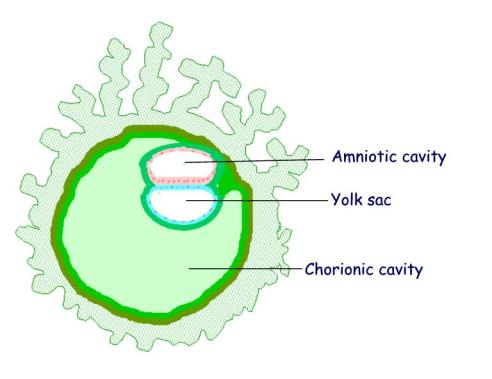
. Placental vertebrate development have both extra-embryonic outside the embryo and intra-embryonic inside the embryo coeloms. Lacunae develop in the mesoderm until they coalesce into a large chorionic cavity surrounding the amniotic and yolk sac cavities. Amniotic fluid is in the amniotic sac.
The extra-embryonic coeloms include the yolk sac amniotic cavity and the chorionic cavity information on these spaces can also be found on. Thus throughout prenatal life humans are surrounded by AF. The amnion arises from the epiblast cells of the blastocyst and grows to surround the developing embryo creating a fluid-filled cavity.
In early gestation two fluid-filled sacs surround the embryo. In this structure the stem cells began to turn into amnion cells organized in a shell around an empty cavity. The amnioticcavity is formedby the fusion of the parts of the amnioticfold which first makes its appearance at the cephalic extremity and subsequently at the caudal end and sides of the embryo.
It is generated from maternal plasma and passes through the fetal membranes by osmotic and hydrostatic forces. Formation of the embryonic disc leaves spaces on either side that develop into the amniotic cavity and the yolk sac. Amniotic Cavity Develops thanks to the proliferation of the epiblast layer of the inner cell mass embryoblast.
In recent years the structure and function of the amnion have been investigated particularly the pluripotent properties of AM cells which are an. It completely surrounds the embryo and delimits the amniotic cavity which is filled by amniotic liquid. It is eventually incorporated into the fetus GI Tract.
Amniotic membrane AM or amnion is a thin membrane on the inner side of the fetal placenta. Within the epiblast a cavity develops the amniotic cavity which fills with amniotic fluid. Cells from the epiblast will also eventually form the body of the embryo.
When fetal kidneys begin to function around week 16 fetal urine also contributes to the fluid. In humans it forms the innermost fetal membrane produces amniotic fluid expanding to eventually fuse with the chorionic membrane during week 8 of development. Where does the amnion come from.
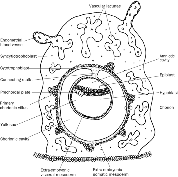
The amniotic sac enlarges rapidly due to an increase in the volume of amniotic fluid. At the beginning of the second week the cells of the inner cell mass form into a two-layered disc of embryonic cells and a spacethe amniotic cavity opens up between it and the trophoblast Figure 5. The chorion is a double-layered membrane formed by the trophoblast and the extra-embryonic mesoderm which eventually will give rise to the fetal part of the placenta.
It develops from a combination of maternal and fetal cells that combine to form the layers of the. Amniotic sac surrounds the fetus and contains the amniotic fluid providing mechanical protection to the developing fetus. The epiblasts can also be called amnion.
This fluid serves many functions critical for prenatal development. How does the amniotic cavity form. Coelom - Greek koilma cavity Term used to describe a fluid-filled cavity or space.
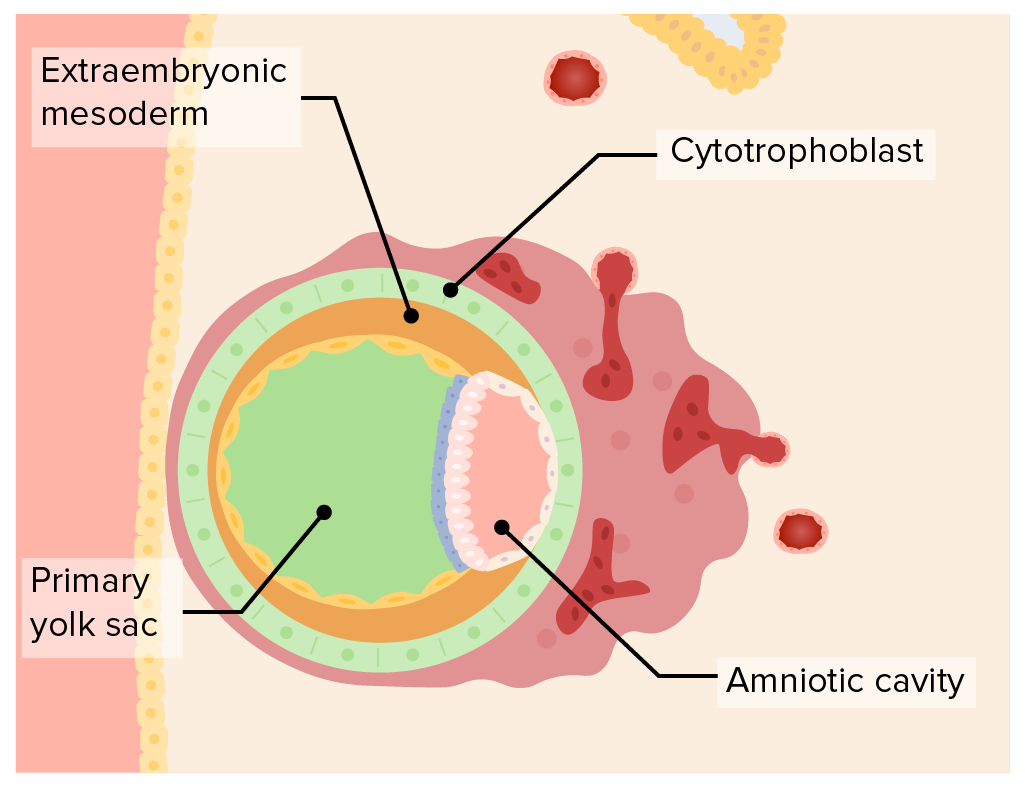
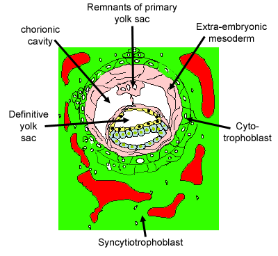
Hypoblast cells migrate to surround the blastocyst cavity differentiate into extraembryonic mesoderm surrounding amniotic and yolk sac cavities. Amniotic fluid is present from the formation of the gestational sac. Extra-embryonic mesoderm Extra-embryonic mesoderm cells migrate between the cytotrophoblast and yolk sac and amnion.
Epiblast Epiblast cells cavitate to form the amnion an extra-embryonic epithelial membrane covering the embryo and amniotic cavity. The amniotic cavity is what contains the amniotic fluid and helps hold the embryo and protect it. The chorion is a double-layered membrane formed by the trophoblast and the extra-embryonic mesoderm which eventually will give rise to the fetal part of the placenta.
A thick soft bed of gel covered with a looser gel which Gumucio compared to a bed of fully congealed Jello with a layer of partially set Jello on top. The team tested different methods to produce amnion. Yolk sac provides nourishment for the fetus.
The amnion lines the amniotic sac and protects the embryo from physical injury. The amniotic cavity is formed by the fusion of the parts of the amniotic fold which first makes its appearance at the cephalic extremity and subsequently at the caudal end and sides of the embryo. The amniotic sac enlarges rapidly due to an increase in the volume of amniotic fluid.
The formation of the coelomic cavity begins during the fourth week of gestation when the exocoelomic cavity splits the extraembryonic mesoderm into the splanchnic mesoderm lining and the somatic mesoderm. The exocoelomic cavity and the amniotic cavity. As the amniotic fold rises and fuses over the dorsal aspect of the embryo the amniotic cavity is formed.
The fluid contained in this sac is the source of. Outline the formation of the uteroplacental circulation. The exocoelomic membrane is derived from the hypoblast and lines the cavity that appears beneath the endoderm the primary yolk sac.
Some epiblast cells become specialized as amnioblasts and they secrete the amniotic fluid. As the amniotic fold rises and fuses over the dorsal aspect of the embryo the amniotic cavity is formed. The amnioticcavity is the closed sacbetween the embryo and the amnion containing the amnioticfluid.
What is the amnion and its function. Primitive Yolk Sac Develops thanks to the proliferation of the hypoblast layer. The amniotic cavity is formed by the fusion of the parts of the amniotic fold which first makes its appearance at the cephalic extremity and subsequently at the caudal end and sides of the embryo.

Extraembryonic Membranes An Overview Sciencedirect Topics

Prenatal Development Amnion Britannica

Cell Intercalation During The Final Step Of Pro Amniotic Cavity Download Scientific Diagram

Embryonic Development Anatomy And Physiology Ii
The Second Week Of Life

Prenatal Development Amnion Britannica
2
Embryoblast And Trophoblast Development Concise Medical Knowledge
Second Week Days 8 14
Implantation And Placentation Clinical Gate

Embryo 2 3 2nd Week Flashcards Quizlet

Amniotic Egg 4 Extra Membranes Amnion Yolk Sac Chorion Allantois Amniotic Cavity Shell Albumin Basic Algebra Female Reproductive System Anatomy Cavities

Remodelling Of Embryo Architecture And Pro Amniotic Cavity Formation A Download Scientific Diagram

Embryonic Development Anatomy And Physiology Ii
Female Reproductive System The Histology Guide

Embryonic Development Anatomy And Physiology Ii

Amniotic Sac Wikiwand

Development Of The Fetal Membranes Osmosis

General Embryology 2nd Week Flashcards Quizlet


Comments
Post a Comment